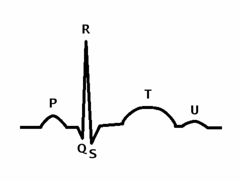
The parts of a QRS complex. Ventricular systole begins at the QRS.
The bulk flow of blood throughout the body is driven by pressure gradients created by the pumping action of the heart. A vast network of blood vessels includes arteries, arterioles, capillaries, venules and veins. This network has two branches, the pulmonary circulation, which carries blood to and from the lungs and the systemic circulation, which delivers blood to the cells throughout the body and and removes wastes for excretion. While at any moment only about 5% of the total blood volume is flowing through the capillaries, it is in the capillaries that the main functions of the circulatory system take place, which are the provision of nutrients to the cells and the removal of metabolic end products. The overall purpose of the circulatory system is to provide adequate blood flow to the capillaries.
While physics underlies biological science in a general way, especially through chemistry, there are some topics where specific physics concepts are directly relevant in a particularly straightforward way. These topics become favorite subjects for a certain type of MCAT passage where the direct application of basic physics concepts allows those concepts to be tested within the biological context. The application of geometric optics concepts to the human eye is an example, and likewise, with the cardiovascular system, the application of fluid mechanics concepts. This means that the cardiovascular system will not only be important for the exam in terms of physiological homeostasis but also for Bernoulli's principle and Poiseuille's law.
WikiPremed Resources
The Cardiovascular System Images
Conceptual Vocabulary Self-Test
Basic Terms Crossword Puzzle
Basic Puzzle Solution
Conceptual Vocabulary for The Cardiovascular System
Blood vessels are part of the cardiovascular system and function to transport blood throughout the body, the most important types being arteries and veins.
An artery is a muscular blood vessel that carries blood away from the heart.
Capillaries are the smallest of a body's blood vessels, connecting arterioles to venules
A vein is a blood vessel that carries blood toward the heart.
The heart is a muscular organ responsible for pumping blood through the blood vessels by repeated, rhythmic contractions.
An arteriole is a small diameter blood vessel that extends and branches out from an artery and leads to capillaries.
A venule is a small blood vessel that allows deoxygenated blood to return from the capillary beds to the larger blood vessels called veins.
The aorta is the largest artery in the human body.
The right atrium is one of four chambers in the human heart, receiving de-oxygenated blood from the superior and inferior vena cavae and the coronary sinus and pumping it into the right ventricle through the tricuspid valve.
The right ventricle is one of four chambers in the human heart, receiving de-oxygenated blood from the right atrium via the tricuspid valve and pumping it into the pulmonary artery via the pulmonary valve.
The left atrium is one of the four chambers in the human heart, receiving oxygenated blood from the pulmonary veins and pumping it into the left ventricle.
The heart valves maintain the unidirectional flow of blood by opening and closing depending on the difference in pressure on each side.
The left ventricle is one of four chambers in the human heart, receiving oxygenated blood from the left atrium via the mitral valve and pumping it into the aorta via the aortic valve.
The pulmonary arteries carry blood from the heart to the lungs.
The tricuspid valve is on the right side of the heart, between the right atrium and the right ventricle.
The mitral valve, also known as the bicuspid valve, is a dual flap valve in the heart that lies between the left atrium and the left ventricle.
Cardiac cycle is the term referring to all or any of the events related to the flow of blood that occur from the beginning of one heartbeat to the beginning of the next.
Heart rate is a term used to describe the frequency of the cardiac cycle.
Hypertension is a medical condition in which the blood pressure is chronically elevated.
The sinoatrial node is the impulse generating pacemaker tissue located in the right atrium of the heart, and thus the generator of sinus rhythm.
The atrioventricular node is an area of specialized tissue between the atria and the ventricles of the heart, which conducts the normal electrical impulse from the atria to the ventricles.
The bundle of His is a collection of heart muscle cells specialized for electrical conduction that transmits the electrical impulses from the AV node to the point of the apex of the fascicular branches.
Purkinje fibers are specialized myocardial fibers located in the inner ventricular walls of the heart that conduct an electrical stimulus or impulse that enables the heart to contract in a coordinated fashion.
The endothelium is the thin layer of cells that line the interior surface of blood vessels.
The hepatic portal vein drains blood from the digestive system and its associated glands.
A portal venous system occurs when a capillary bed drains into another capillary bed through veins.
The pulmonary veins carry oxygen-rich blood from the lungs to the left atrium of the heart.
The aortic valve is one of the valves of the heart. It lies between the left ventricle and the aorta.
The superior and inferior vena cavae are the veins that return de-oxygenated blood from the body into the heart, emptying into the right atrium.
Systole is the contraction of heart chambers, driving blood out of the chambers.
Diastole is the period of time when the heart relaxes after contraction.
Vascular resistance is a term used to define the resistance to flow that must be overcome to push blood through the circulatory system.
A vasoconstrictor, also vasopressor or simply pressor, is any substance that acts to cause vasoconstriction and usually results in an increase of the blood pressure.
Peripheral arteries are the arteries which are furthest from the heart.
A sinusoid is a small blood vessel similar to a capillary but with a discontinuous endothelium.
Hypotension refers to an abnormally low blood pressure.
Sinus rhythm is a term used in medicine to describe the normal beating of the heart, as measured by an electrocardiogram (ECG).
A vasodilator is a drug or chemical that relaxes the smooth muscle in blood vessels, which causes them to dilate.
The cardiac action potential is a specialized action potential in the heart, with unique properties necessary for function of the electrical conduction system of the heart.
The tunica externa, previously known as the tunica adventitia, is the outermost layer of a blood vessel, surrounding the tunica media.
The tunica media is the middle layer of an artery or vein
The tunica intima is the innermost layer of an artery.
Superficial vein is a term used to describe a vein that is close to the surface of the body.
Myocardium is the muscular tissue of the heart.
Atrial fibrillation is a cardiac arrhythmia that involves the two atria of the heart.
A electrocardiogram (ECG or EKG) is a graphic produced by an electrocardiograph, which records the electrical activity of the heart over time.
Cardiac output is the volume of blood being pumped by the heart, in particular by a ventricle in a minute.
Stroke volume is the amount of blood pumped by the left ventricle of the heart in one contraction.
End-systolic volume is the volume of blood in the ventricles just after systole.
End-diastolic volume is the volume of blood in a ventricle at the end of filling.
The baroreflex or baroreceptor reflex is one of the body's homeostatic mechanisms for maintaining blood pressure, in which an elevated blood pressure reflexively causes blood pressure to decrease.
The jugular veins are veins that bring deoxygenated blood from the head back to the heart via the superior vena cava.
The superior vena cava is a large, short vein that carries de-oxygenated blood from the upper half of the body to the heart's right atrium.
The inferior vena cava is the large vein that carries de-oxygenated blood from the lower half of the body into the heart.
The interventricular septum is the stout wall separating the ventricles of the heart from one another.
The coronary sinus is a collection of veins joined together to form a large vessel that collects blood from the myocardium of the heart.
Chronotropic effects are those that change the heart rate.
The systemic venous system describes the veins that drain into the right atrium without passing through two vascular beds before reaching the right side of the heart.
The endocardium is the innermost layer of tissue that lines the chambers of the heart.
Epicardium describes the outer layer of heart tissue.
Preload is the pressure stretching the ventricle of the heart, after passive filling and atrial contraction.
Starling's law states that the more the ventricle is filled with blood during diastole, the greater the volume of ejected blood will be during the resulting systolic contraction.
A dromotropic agent is one which affects the conduction velocity of the AV node, and subsequently the rate of electrical impulses in the heart.

WikiPremed is a trademark of Wisebridge Learning Systems LLC. The work of WikiPremed is published under a Creative Commons Attribution NonCommercial ShareAlike License. There are elements of work here, such as a subset of the images in the archive from WikiPedia, that originated as GNU General Public License works, so take care to follow the unique stipulations of that license in printed reproductions. |