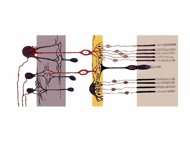
Axial organization of the retina.
Sensory systems are part of the nervous system. Sensory systems transduce stimuli received from the external world (or from within the body) into neural signals that lead to our perceptions of the world. A sensory system consists of sensory receptors, the neural pathways that conduct signals from the receptors to the brain, and the structures in the brain which are dedicated to processing the information.
With so much overlap from other areas of biological science as well as within physical science, sensory systems really are ideal subject matter for MCAT passages. There is also a significant body of knowledge involving the physiology of sensory systems within the field of psychology. There is a separate section on this site dealing with the particular issues regarding sensation and perception from the point of view of psychology.
WikiPremed Resources
Sensory Systems Images
Conceptual Vocabulary Self-Test
Basic Terms Crossword Puzzle
Basic Puzzle Solution
Conceptual Vocabulary for Sensory Systems
A stimulus is a detectable change in the internal or external environment.
Taste or gustation is a form of direct chemoreception producing the ability to detect the flavor of substances such as food and poisons.
Olfaction refers to the sense of smell.
Hearing (or audition) refers to the ability to detect sound.
The tympanic membrane, colloquially known as the eardrum, is a thin membrane that separates the external ear from the middle ear.
The cochlea is the auditory portion of the inner ear.
Eyes are organs of vision that detect light.
The cornea is the transparent front part of the eye that covers the iris, pupil, and anterior chamber, providing most of an eye's optical power.
A sensory receptor is a structure that recognizes a stimulus in the internal or external environment, and in response, initiates sensory transduction by creating graded potentials or action potentials in the same cell or in an adjacent one.
The vestibular system, or balance system, is the sensory system that provides the dominant input about our movement and orientation in space.
Hair cells are the sensory receptors of both the auditory system and the vestibular system in all vertebrates.
The middle ear is the portion of the ear internal to the eardrum, and external to the oval window of the cochlea.
The Eustachian tube is a tube that links the pharynx to the middle ear.
The ossicles are the three smallest bones in the human body, contained within the middle ear and serving to transmit sounds to the fluid-filled cochlea.
The incus is the anvil-shaped small bone or ossicle in the middle ear.
The stapes is the stirrup-shaped small bone or ossicle in the middle ear which attaches the incus to the fenestra ovalis.
The malleus is a hammer-shaped small bone or ossicle of the middle ear which connects with the incus and is attached to the inner surface of the eardrum.
The inner ear is the bony labyrinth, a system of passages comprising two main functional parts: the cochlea and the vestibular apparatus.
The organ of Corti, or spiral organ, is the organ in the inner ear of mammals that contains auditory sensory cells, or hair cells.
The iris consists of pigmented fibrovascular tissue known as a stroma, which connects a sphincter muscle to contract the pupil, and a set of dilatory muscles to open it.
The retina is a thin layer of neural cells that lines the back of the eyeball.
Rod cells are photoreceptor cells in the retina of the eye that can function in less intense light than cone cells can.
A light-sensitive derivative of vitamin A, retinal is the fundamental chromophore involved in the transduction of light into visual signals.
Accommodation is the process by which the eye increases optical power to maintain a clear image of an object as it draws near the eye.
A mechanoreceptor is a sensory receptor that responds to mechanical pressure or distortion.
A thermoreceptor is a sensory receptor that responds to temperature, primarily within the innocuous range.
Baroreceptors detect the pressure of blood flowing through them, and can send messages to the central nervous system to increase or decrease total peripheral resistance and cardiac output.
A nociceptor is a sensory receptor that sends signals that cause the perception of pain in response to potentially damaging stimulus.
The gustatory system is the sensory system that uses taste buds on the upper surface of the tongue to provide information about the taste of food being eaten.
An olfactory receptor neuron is the primary transduction cell in the olfactory system.
The labyrinth is a system of fluid passages in the inner ear, including both the cochlea which is part of the auditory system, and the vestibular system which provides the sense of balance.
The semicircular canals are three half-circular, interconnected tubes located inside each ear that are the equivalent of three gyroscopes located in three orthogonal planes.
Otoliths are small particles, composed of a combination of a gelatinous matrix and calcium carbonate in the viscous fluid of the saccule and utricle.
Perilymph is an extracellular fluid located within the scala tympani and scala vestibuli of the cochlea.
Endolymph is the fluid contained in the membranous labyrinth of the inner ear.
Also called the vestibular window, the oval window is a membrane-covered opening which leads from the middle ear to the vestibule of the inner ear.
The round window is one of two openings along with the oval window that connect the inner ear to the middle ear.
The ciliary body is the circumferential tissue inside the eye composed of the ciliary muscle and ciliary processes.
Rhodopsin, or visual purple, is a pigment consisting of an opsin protein and retinal cofactor that is responsible for both the formation of the photoreceptor cells and the perception of light.
Opsins are a group of light-sensitive membrane-bound G protein-coupled receptors of the retinylidene protein family found in photoreceptor cells of the retina.
The visual cycle is the biological conversion of a photon into an action potential in the retina.
Cone cells are photoreceptor cells in the retina of the eye which function best in relatively bright light.
A chemosensor, also known as chemoreceptor, is a cell or group of cells that transduce a chemical signal into an action potential.
An osmoreceptor is a sensory receptor primarily found in the hypothalamus of most homeothermic organisms that detects changes in osmotic pressure.
Proprioception is the sense of the relative position of neighbouring parts of the body.
The olfactory epithelium is a specialized epithelial tissue inside the nasal cavity that is involved in smell.
Pacinian corpuscles are nerve endings in the skin, responsible for sensitivity to deep pressure touch and high frequency vibration.
Meissner's corpuscles (or tactile corpuscles) are a type of mechanoreceptor responsible for sensitivity to light touch.
Merkel nerve endings are mechanoreceptors found in the skin in which each ending consists of a cell in close apposition with an enlarged nerve terminal.
Merkel cells are large oval cells found in the skin of vertebrates associated with the sense of touch.
The bony labyrinth ocated in the inner ear consists of the vestibule, the semicircular canals, and the cochlea.
The utricle along with the saccule is one of the two otolith organs located in the vertebrate inner ear.
The membranous labyrinth is lodged within the bony labyrinth, having the same general form, partly separated from the bony walls by a quantity of perilymph.
The ciliary muscle is a smooth muscle responsible for accommodation of the eye.
Also known as iodopsins, photopsins are the photoreceptor proteins found in the cone cells of the retina that are the basis of color vision.
The Ruffini ending or corpuscle is a class of slowly adapting mechanoreceptor thought to exist only in the glabrous dermis and subcutaneous tissue of humans
The fovea is a part of the eye, located in the center of the macula region of the retina, which is responsible for sharp central vision
Photoisomerization is structural change between isomers is caused by photoexcitation.
A free nerve ending is an unspecialized, afferent nerve ending, which are are unencapsulated and have no complex sensory structures.
The Golgi tendon organ is a proprioceptive sensory receptor organ that is located at the insertion of skeletal muscle fibres into the tendons of skeletal muscle.
The saccule is the smaller of the two vestibular sacs.
The basilar membrane within the cochlea of the inner ear is a stiff structural element that separates the two liquid-filled tubes that run along the coil of the cochlea.
The sclera is the opaque, usually white, fibrous, protective layer of the eye containing collagen and elastic fibers.
The choroid is the vascular layer of the eye lying between the retina and the sclera.
The shape theory of smell states that the sensation of smell is due to a 'lock and key' mechanism by which a scent molecule fits into olfactory receptors in the nasal lamina of the nose.
The receptive field of a sensory neuron is a region of space in which the presence of a stimulus will alter the firing of that neuron.
The vestibule is the central part of the osseous labyrinth, situated medial to the tympanic cavity, behind the cochlea, and in front of the semicircular canals.
The uvea, also called the vascular tunic, is the pigmented middle of the three concentric layers that make up an eye.
A kinocilium is a special structure connected to the hair cells of the inner ear's cochlea which acts to aid in depolarization and hyperpolarization of the plasma membrane due to bending of sterocillia.
The optic disc is the location where ganglion cell axons exit the eye to form the optic nerve.
C-fibers are unmyelinated nerve fibers associated with chronic or dull pain.
The macula is an oval yellow spot near the center of the retina of the human eye.
The retinal pigment epithelium is the pigmented cell layer just outside the neurosensory retina that nourishes retinal visual cells.
As a part of the retina, bipolar cells exists between photoreceptors and ganglion cells.
Amacrine cells are interneurons in the retina which deliver 70% of the ganglion cells input, and also regulate the output of the cone bipolar cells which deliver the other 30%.
The fungiform papillae are mushroom shaped papillae on the tongue.
The foliate papillae is a localized area at the side of the base of the tongue in which taste buds are especially abundant.
The spiral ganglion is the group of nerve cells that serve the sense of hearing by sending a representation of sound from the cochlea to the brain.
The sclera and cornea form the fibrous tunic of the bulb of the eye.
The zonule of Zinn is a ring of fibrous strands connecting the ciliary body with the crystalline lens of the eye.
Horizontal cells are the laterally interconnecting neurons in the outer plexiform layer of the retina.

WikiPremed is a trademark of Wisebridge Learning Systems LLC. The work of WikiPremed is published under a Creative Commons Attribution NonCommercial ShareAlike License. There are elements of work here, such as a subset of the images in the archive from WikiPedia, that originated as GNU General Public License works, so take care to follow the unique stipulations of that license in printed reproductions. |