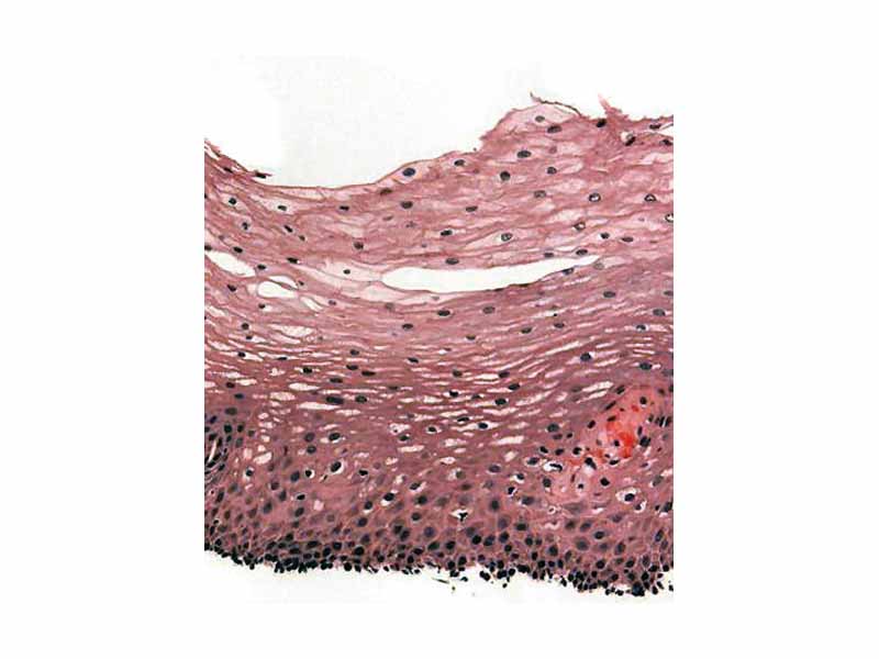
H&E; stain of biopsy of normal esophagus showing the stratified squamous cell epithelium.
The single unifying principle for the science sections of the new MCAT is a levels of organization approach to living systems that begins with the chemistry underlying the behavior of macromolecules and extends through the cell all the way to organ systems and physiological homeostasis. Biological tissues represent a cellular organizational level intermediate between cells and the organ systems of physiology. As a subject matter for MCAT review the tissue level doesn't have as large a conceptual vocabulary as cell biology or physiology, but it is very important. At least a basic understanding of the structure of tissues lends a unifying coherence to both cell biology and physiology.
Additionally, there is an important body of lab techniques associated with the study of tissues, namely, the techniques of histology. Histology is the art of preparing and visualizing tissue samples for microscopy, ie. the techniques of fixation, embedding, sectioning, clearing, staining and mounting. Histology represents a core laboratory practice, and you need the basics for the exam.
WikiPremed Resources
Animal Development and Embryology Images
Conceptual Vocabulary Self-Test
Basic Terms Crossword Puzzle
Basic Puzzle Solution
Conceptual Vocabulary for Tissues and Histology
Fertilization is fusion of gametes to form a new organism of the same species.
Mesoderm is a primary germ layer which forms during gastrulation when some of the cells migrating inward to form the endoderm form an additional layer between the endoderm and the ectoderm.
Endoderm is one of the primary germ layers formed during animal embryogenesis when cells migrating inward along the archenteron form the inner layer of the gastrula.
The ectoderm is the primary germ layer which emerges first during embryogenesis and forms from the outermost of the germ layers.
A germ layer is a collection of cells, a primary tissue layer, formed during animal embryogenesis.
The blastula stage of embryonic development in animals follows the morula and precedes the gastrula stage in the developmental sequence.
A zygote is the cell that results from fertilization.
A blastopore is an opening into the archenteron during the embryonic stages of an organism.
The notochord is a flexible, rod-shaped body found in embryos of all chordates which is composed of cells derived from the mesoderm and defines the primitive axis of the embryo.
A morula is an embryo at an early stage of embryonic development, consisting of approximately 12-32 cells in a solid ball contained within the zona pellucida.
Organogenesis is the process in animal development by which the ectoderm, endoderm, and mesoderm develop into the internal organs of the organism.
Cleavage is the division of cells in the early embryo.
The blastocyst is the structure formed in early human embryogenesis, after the formation of the blastocele, but before implantation, possessing an inner cell mass, or embryoblast, and an outer cell mass, or trophoblast.
Gastrulation is a phase early in the development of animal embryos, during which the morphology of the embryo is dramatically restructured by cell migration.
The gastrula phase of embryonic development, which follows after the blastula stage, is seen in all animals except the sponges.
The archenteron is known as the primitive gut that forms during gastrulation in the developing blastula.
Blastomere is the term for the cells formed by cleavage of the ovum in very early embryonic development.
Following primary and secondary neurulation, the neural tube is the developing vertebrate embryo's precursor to the central nervous system.
Formation of neural plate is the first step of neurulation in human embryology. It is created by a flat thickening opposite to the primitive streak.
A blastocoele is the fluid-filled central region of a blastocyst.
Embryogenesis is the process by which the embryo is formed and develops.
In early embryogenesis, the inner cell mass is the mass of cells inside the primordial embryo that will eventually give rise to the definitive structures of the fetus.
Trophoblasts are cells forming the outer layer of a blastocyst, which provide nutrients to the embryo and develop into a large part of the placenta.
The dorsal nerve cord is one of the embryonic features unique to chordates, along with a notochord, a post-anal tail and pharyngeal slits.
The primitive streak is a structure that forms during the early stages of embryonic development, characterized as a furrow in the midline of the embryonic disk at the future caudal end of the embryo.
During the early stages of embryonic development, a shallow groove, the primitive groove, appears on the surface of the primitive streak
The primitive knot is the organizer for gastrulation in vertebrates, starting as a regional knot of cells that forms on the blastodisc immediately anterior to where the outer layer of cells will begin to migrate inwards.
Angiogenesis is a physiological process involving the growth of new blood vessels from pre-existing vessels.
The term vegetal pole refers to the hemisphere of a blastula embryo which contains large yolky cells that divide very slowly.
The term animal pole refers to the hemisphere of a blastula embryo which consists of small cells that divide rapidly, in contrast with the vegetal pole.
Epiboly is the expansion of one cell sheet over other cells. Takes place during gastrulation.
Mesenchyme is the mass of tissue that develops mainly from the mesoderm of an embryo which contains collagen bundles and fibroblasts and later differentiates into blood vessels, blood-related organs, and connective tissues.
Neurulation is the part of organogenesis in vertebrate embryos that includes the events from the formation of the dorsal nerve cord to the eventual formation of the central nervous system.
During primary neurolation, the neural folds make their appearance in front of the primitive streak as two longitudinal ridges, caused by a folding up of the ectoderm.
A morphogen is a substance governing the pattern of tissue development and, in particular, the positions of the various specialized cell types within a tissue.
Somites are masses of mesoderm in the developing vertebrate embryo distributed along the two sides of the neural tube and that will eventually become dermis, skeletal muscle, and vertebrae.
Carnegie stages are a standardized system of 23 stages used in embryology to provide a unified developmental chronology of the vertebrate embryo.
Diploblasty is a condition of the ovum in which there are two primary germ layers: the ectoderm and endoderm, such as with cnidaria and ctenophores.
Triploblasty is a condition of the blastula in which there are three primary germ layers: the ectoderm, mesoderm, and endoderm, such as in higher and intermediate animals from flat worms to humans.
Convergent extension is the process during organogenesis in which layers of cells intercalate and become longer.
Spiral cleavage is the type type of holoblastic cleavage in embryonic development in which at the third cleavage the halves are oblique to the polar axis and typically produce an upper quartet of smaller cells that come to be set between the furrows of the lower quartet.
Nucleus pulposus is the jelly-like substance in the middle of the spinal disc which is the remnant of the notochord.
Paraxial mesoderm is the area of mesoderm that forms just lateral to the neural tube on both sides.
In the developing nervous system, the floor plate is a neural tube structure that separates the left and right components of the basal plate.
The alar plate is a neural structure in the embryonic nervous system, part of the dorsal side of neural tube, that involves the communication of general somatic and general visceral sensory impulses.
A rhombomere is a transiently divided segment in the vertebrate embryo of the developing neural tube in the area that will eventually become the rhombencephalon.
The cephalic flexure is the first flexure, or bend, of the embryonic brain; it appears in the region of the mid-brain.
The myelencephalon is a developmental categorization of a portion of the central nervous system composed of the medulla oblongata and containing a portion of the fourth ventricle and various cranial nerves.
The metencephalon is a developmental categorization of portions of the central nervous system composed of the pons, the cerebellum, a portion of the fourth ventricle, and various cranial nerves.
Isolecithal refers to the even distribution of yolk in the cytoplasm of ovums of mammals and other invertebrates.
Centrolecithal is the placement of the yolk in the centre of the cytoplasm of ovums such as within many arthropod eggs.
Chordin is a polypeptide that dorsalizes the developing embryo by binding ventralizing proteins such as bone morphogenetic proteins.
Bone morphogenetic proteins are a group of growth factors and cytokines known for their ability to induce the formation of bone and cartilage.
Noggin is a polypeptide that binds to members of the TGF-beta superfamily of proteins. It is a Bone morphogenetic protein inhibitor.
The transforming growth factor beta superfamily includes inhibins, activin, anti-mû¥llerian hormone, bone morphogenetic protein, decapentaplegic and Vg-1.
The hedgehog signaling pathway is one of the key regulators of animal development conserved from flies to humans. It is involved in establishing the basis of the body plan.
Sonic hedgehog homolog is one of three proteins in the mammalian hedgehog family which play a key role in regulating vertebrate organogenesis, the others being desert hedgehog and Indian hedgehog.
The Wnt signaling pathway describes a complex network of proteins most well known for their roles in embryogenesis and cancer, but also involved in normal physiological processes in adult animals.
Epidermal growth factor or EGF is a growth factor that plays an important role in the regulation of cell growth, proliferation and differentiation.
Fibroblast growth factors, or FGFs, are a family of growth factors involved in wound healing and embryonic development.
In vertebrate embryonic development, a myotome is a group of tissues formed from somites that develop into the body wall muscle.
A myoblast is a type of stem cell that exists in muscles.
A sclerotome is part of a somite, a structure in vertebrate embryonic development, which eventually differentiate into the vertebrae and most of the skull.
The somitomeres are loose masses of paraxial mesoderm derived cells in the developing vertebrate embryo that form along each side of the neural tube towards the end of the third gestational week.
Essential for mesoderm formation and neural tube formation, epithelial-mesenchymal transition is a program of development characterized by loss of cell adhesion, repression of E-cadherin expression, and increased cell mobility.
Agrin is a large proteoglycan with a role in the development of neuromuscular junctions during embryogenesis.

WikiPremed is a trademark of Wisebridge Learning Systems LLC. The work of WikiPremed is published under a Creative Commons Attribution NonCommercial ShareAlike License. There are elements of work here, such as a subset of the images in the archive from WikiPedia, that originated as GNU General Public License works, so take care to follow the unique stipulations of that license in printed reproductions. |