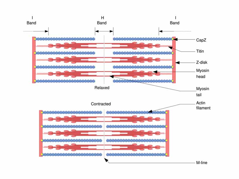
Sliding filament model of muscle contraction.
Consisting of muscles, ligaments, tendons, bones and cartilage, the musculoskeletal system supports the body, provides for motion, and protects vital organs. Muscle cells are specialized to use chemical energy to produce mechanical force. Three types of muscle can be identified: skeletal muscle, smooth muscle, and cardiac muscle. The other primary tissue type within the musculoskeletal system is bone, a specialized connective tissue consisting of a collagenous matrix impregnated with calcium phosphate and other minerals. Bones serve as the main storage system for calcium and phosphorus. Bones also contain critical components of the hematopoietic system.
WikiPremed Resources
The Musculoskeletal System Images
Conceptual Vocabulary Self-Test
Basic Terms Crossword Puzzle
Basic Puzzle Solution
Conceptual Vocabulary for The Musculoskeletal System
A muscle fiber is a single cell of a muscle.
Muscle is contractile tissue of the body and is derived from the mesodermal layer of embryonic germ cells.
Myosins are a large family of motor proteins found in eukaryotic tissues which are responsible for actin-based motility.
Actin is the monomeric subunit of microfilaments, one of the three major components of the cytoskeleton, and of thin filaments which are part of the contractile apparatus in muscle cells.
Smooth muscle is a type of non-striated muscle, found within the bladder, abdominal cavity, the uterus, male and female reproductive tracts, the gastrointestinal tract and elsewhere.
Cardiac muscle is a type of involuntary striated muscle found within the heart.
Bones are rigid organs that form part of the endoskeleton of vertebrates, functioning to move, support, and protect the various organs of the body, produce red and white blood cells and store minerals.
Skeletal muscle is a type of striated muscle, usually attached to the skeleton.
Myofibrils are cylindrical organelles, found within muscle cells, which are bundles of actomyosin filaments that run from one end of the cell to the other, attached to the cell surface membrane at each end.
A sarcomere is the basic unit of a muscle's cross-striated myofibril.
A muscle contraction occurs when a muscle fiber generates tension through the action of actin and myosin cross-bridge cycling.
As part of the regulation of muscle contraction, in resting muscle fibres, the protein tropomyosin is displaced from its normal binding groove by troponin.
Bone marrow is the soft tissue found in the hollow interior of bones.
Osseous tissue forms the rigid part of the bone organs that make up the skeletal system.
An osteoblast is a mononucleate cell that is responsible for bone formation.
An osteoclast is a type of bone cell that removes bone tissue by removing the bone's mineralized matrix.
Cartilage is a type of dense connective tissue composed of collagen fibers and/or elastin fibers which can supply smooth surfaces for the movement of articulating bones.
A tendon or sinew is a tough band of fibrous connective tissue that connects muscle to bone and is built to withstand tension.
The sarcoplasmic reticulum is a special type of smooth endoplasmic reticulum found in smooth and striated muscle.
The sarcolemma is the cell membrane of a muscle cell.
A transverse or T-tubule is a deep invagination of the plasma membrane found in skeletal and cardiac muscle cells which allows depolarization of the membrane to quickly penetrate to the interior of the cell.
An intercalated disc is an undulating double membrane separating adjacent cells in cardiac muscle fibers.
The a star-shaped osteocyte is the most abundant cell found in bone, derived from osteoblasts after they become trapped within the matrix they secrete.
Bone resorption is the process by which osteoclasts break down bone and release the minerals, resulting in a transfer of calcium from bone fluid to the blood.
The inorganic mineral hydroxylapatite makes up seventy percent of bone.
The diaphysis is the main or mid section shaft of a long bone.
Chondrocytes are the only cells found in cartilage.
The muscle spindle's functions are to send proprioceptive information about the muscle to the central nervous system and to respond to muscle stretching.
A motor unit is a single alpha-motor neuron and all of the corresponding muscle fibers it innervates.
A myogenic muscle doesn't need to receive impulses from a nerve to make it contract.
Phosphocreatine is a phosphorylated creatine molecule that is an important energy store in skeletal muscle.
Creatine is nitrogenous organic acid which naturally occurs in vertebrates and helps to supply energy to muscle and nerve cells.
Also known as compact bone, cortical bone is dense and forms the surface of bones.
Haversian canals are a series of tubes around narrow channels formed by lamellae in compact bone.
Also known as trabecular, or spongy bone, cancellous bone fills the inner cavity of long bones. It has low density and strength, but very high surface area.
Intramembranous ossification is the type of bone formation responsible for the development of flat bones, especially those found in the skull and clavicles.
Endochondral ossification is the type of bone formation responsible for much of the bone growth in vertebrate skeletons, especially in long bones.
The epiphyseal plate, or growth plate, is the cartilage plate in the long bones of children and adolescents.
A chondroblast is a cell originating from a mesenchymal stem cell which forms chondrocytes or cartilage cells.
a perimysium is a sheath of connective tissue which groups individual muscle fibers into bundles or fascicles.
The endomysium is a layer of connective tissue that ensheaths a muscle fiber and is composed mostly from reticular fibers.
Titin, also known as connectin, is a protein important in the contraction of striated muscle tissues, connecting the Z line to the M line in the sarcomere.
Localized to the I-band of sarcomeres in skeletal muscle, nebulin is a very large protein, binding as many as 200 actin monomers.
Satellite cells are mononuclear progenitor cells found in mature muscle between the basal lamina and sarcolemma, which are able to differentiate and fuse to augment existing muscle fibres and to form new fibres.
Creatine kinase catalyses the conversion of creatine to phosphocreatine.
Epiphysis is the name for a rounded end of a long bone.
Hyaline cartilage consists of a slimy mass of a firm consistency, but of considerable elasticity and pearly bluish color, containing no nerves or blood vessels.
A fascicle is a bundle of skeletal muscle fibers surrounded by connective tissue.
Intrafusal fibers are muscle fibers that comprise the muscle spindle.
In contrast to intrafusal fibers, extrafusal muscle fibers are the class of muscle fiber innervated by alpha motor neurons which generate tension, perform mechanical work and allow for movement by contracting.
Excitation-contraction coupling is a term which describes the physiological process of converting an electrical stimulus to mechanical response.
Motor unit recruitment is the progressive activation of a muscle by successive recruitment of contractile units motor units to accomplish increasing gradations of contractile strength.
Calmodulin (CaM) is a ubiquitous, calcium-binding protein that can bind to and regulate a multitude of different protein targets, thereby affecting many different cellular functions.
Ryanodine receptors form a class of calcium channels which are the major cellular mediators of calcium induced calcium release in animal cells.
Myocardium is the muscular tissue of the heart.
Canaliculi are microscopic canals between the various lacunae of ossified bone into which the radiating processes of the osteocytes project.
A lacuna is a small space containing an osteocyte in bone or chondrocyte in cartilage.
Osteoid is a protein mixture secreted by osteoblasts which mineralizes and becomes bone.
Cathepsin K is a protein with the ability to catabolize elastin, collagen, and gelatin which allows it to break down bone and cartilage.
Elastic cartilage is a type of cartilage present in the outer ear, larynx, and epiglottis which contains fibers made of elastin.
White fibrocartilage consists of a mixture of white fibrous tissue and cartilaginous tissue in various proportions.
The metaphysis is the portion of a long bone between the epiphyses and the diaphysis.
A myoblast is a type of stem cell that exists in muscles which differentiate into satellite cells.
1 to 3 nuclear bag fibers lie in the center of each intrafusal muscle fibre of a muscle spindle, which cause excitation of both the primary and secondary nerve fibres.
A nuclear chain fiber is a specialized sensory organ contained within a muscle, which, along with nuclear bag fibers, make up the muscle spindle responsible for the detection of changes in muscle length.
Dystrophin is a rod-shaped cytoplasmic protein, and a vital part of a protein complex that connects the cytoskeleton of a muscle fiber to the surrounding extracellular matrix through the cell membrane.
A tetanic contraction occurs when a motor unit has been maximally stimulated by its motor neuron.
The Interstitial cell of Cajal is a type of cell found in the gastrointestinal tract that serves as a pacemaker that triggers gut contraction.
The endosteum is a thin layer of connective tissue which lines the surface of the bony tissue that forms the medullary cavity of long bones.
The medullary cavity is the central cavity of bone shafts where yellow marrow is stored.
The periosteum is a thin layer of dense, irregular connective tissue membrane that covers the outer surface of a bone in all places except at joints.
Enthesis is the point at which a tendon inserts into bone, where the collagen fibers are mineralized and integrated into bone tissue.
A type Ia sensory fiber also called primary afferent fiber is a component of a muscle fiber's muscle spindle which keeps track of the how fast a muscle stretch changes.
Type II sensory fibers are the second of the two main groups of stretch receptors. Their firing rate is directly related to the muscle's instantaneous length, or position.
Calsequestrin is a calcium-binding protein of the sarcoplasmic reticulum.
The costameres are sub-sarcolemmal protein assemblies circumferentially aligned in register with the Z-disk of peripheral myofibrils.
Dystrobrevin is a protein that binds to dystrophin in the costamere of skeletal muscle cells.
Dysbindin, short for dystrobrevin-binding protein 1, is a protein constituent of the dystrophin-associated protein complex of skeletal muscle cells.
The sarcoglcyans are a family of five transmembrane proteins involved in the protein complex responsible for connecting the muscle fibre cytoskeleton to the extracellular matrix.
A triad is a histological structure formed by a T tubule with a sarcoplasmic reticulum cisterna on either side.
Myosin-light-chain kinase is a serine/threonine-specific protein kinase which phosphorylates myosin.
Fascia adherens is broad intercellular junction in the intercalated disk of cardiac muscle anchoring actin filaments.
Sharpey's fibers are a matrix of connective tissue consisting of bundles of strong collagenous fibres connecting periosteum to bone.
The perichondrium is a layer of dense irregular connective tissue which surrounds the cartilage of developing bone.

WikiPremed is a trademark of Wisebridge Learning Systems LLC. The work of WikiPremed is published under a Creative Commons Attribution NonCommercial ShareAlike License. There are elements of work here, such as a subset of the images in the archive from WikiPedia, that originated as GNU General Public License works, so take care to follow the unique stipulations of that license in printed reproductions. |